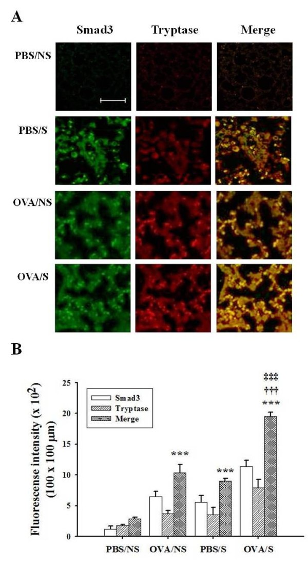
Figure 7.

Effects of smoke exposure on co-localization of mast cell tryptase and Smad3 in lung tissues of OVA-induced asthmatic mice. Experimental conditions and group symbols used were described in Fig. 1. Lung tissues were fixed with 4% paraformaldehyde, sectioned and immunostained as described in "Materials and Methods". Co-localization was examined by immunohistochemistry (A). The degree of IHC color (yellow) developed by co-localization was quantified by intensity in 100 × 100 μm areas under microscopy (5 areas/each slide × 8 mice/each group = 40 areas), and then mean ± SEM for 40 areas was presented by histogram (B). ***, P<0.001 versus PBS/NS mice. †††, P < 0.001 versus OVA/NS mice. ‡‡‡, P < 0.001 versus PBS/S mice. Bar in control (PBS/NS) image indicates 50 μm.
