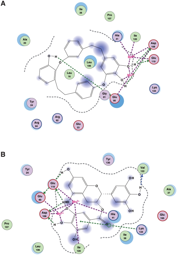
Figure 8. Comparison between marchantins and catechins that bind to PA endonuclease.
Docking simulation analysis of marchantin A (panel A) or EGCG (panel B) with the PA endonuclease domain. Two dimensional analysis of the interactions of marchantin A or EGCG with PA endonuclease is shown. The chemical structures of marchantin A and EGCG is shown in the center. The interacting amino acids of PA endonuclease are shown around them. The dihydroxyphenyl groups of both marchantin A and EGCG interact with two manganese ions.

