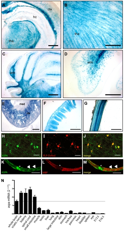
Figure 2. Analysis of aspa expression in CNS and periphery.
A–D, Representative pictures of β-Galactosidase staining in adult aspalacZ/+ mouse tissues show activity of the aspa gene in oligodendrocytes of white and grey matter throughout the brain. Abundant staining was detected in white matter tracts in cerebellum and corpus callosum. While in the hippocampus and cortex X-Gal staining was found in oligodendrocyte cell bodies (A), lacZ-positive fibres were detected in the thalamus (B). In the cerebellum, proximal processes and somata of white matter oligodendrocytes were stained (C, D). E–F, Outside the CNS, the cortex of the kidney (E), small intestine (F) and sciatic nerve fibres (G) show intense X-Gal staining. H–J, Confocal detection of ASPA immunoreactivity (H) in cerebellar white matter of an adult Plp-DsRed-1 transgenic mouse expressing the red fluorescent reporter protein (I) in oligodendrocytes [15]. ASPA is expressed in somata and processes of oligodendrocytes. The pattern of ASPA-immunopositive cells matches the DsRed-expressing oligodendrocytes (J). K–M, Confocal detection of ASPA immunoreactivity in cytosolic domains of wildtype Schwann cells. ASPA is enriched at the paranode (asterisk) and bands of Cajal (arrow heads) and segregates from MBP. N, Q-PCR analysis of aspa mRNA levels in different tissues of adult aspa+/+ mice (n = 6). hc, hippocampus; ctx, cortex; med, medulla; tha, thalamus; wm, white matter. Bars: 500 µm in A,C,E; 200 µm in B, D; 400 µm in F,G; 20 µm in H–J; 10 µm in K–M.

