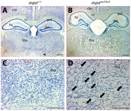
Figure 3. CNS vacuolisation in aspalacZ/lacZ mutants.
Representative pictures of Nissl stained brain sections of aspa+/+ (A,C) and aspalacZ/lacZ (B,D) mice (4 months). Vacuoles are abundant in forebrain grey matter of cortex and thalamus while white matter of the corpus callosum is spared (B). In the hippocampus, the histopathology is exclusively seen in the pyramidal but not the dentate granule cell layer. Magnifications of thalamic areas show substantial spongy degeneration in the mutant (arrows in D) but not in the control (C). hc, hippocampus; cc, corpus callosum; ctx, cortex; dcl, granule cell layer; pcl, pyramidal cell layer; tha, thalamus. Bars: 400 µm in A,B; 200 µm in C,D.

