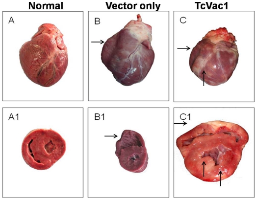
Figure 6. Morphological alterations in the heart.
Dogs were vaccinated and infected with T. cruzi as above. Shown are representative morphologic alterations of the heart during the acute infection phase (60 dpi) in dogs injected with vector only (B&B1) or immunized with TcVac1 (C&C1). Images of normal heart (A&A1) are shown for comparison. Horizontal arrows show right ventricle wall thinning characteristic of ventricle dilation. Vertical arrows show pale striated epicardium and myocardium, characteristic of necrosis produced after inflammatory response to infection.

