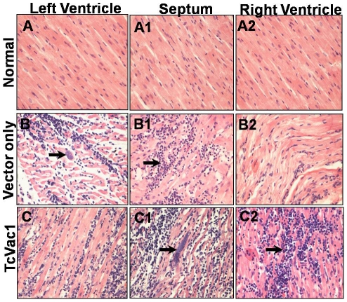
Figure 7. Histological analysis of hearts.
Dogs were vaccinated, and challenged with T. cruzi. Heart tissue sections (5-ìM) from left ventricle, septum, and right ventricle were obtained at 60 days post-infection (acute phase), and stained with hematoxylin-eosin. Shown are representative micrographs of dogs injected with vector only (B,B1,B2) or immunized with TcVac1 (C,C1,C2). Micrographs from normal/uninfected dogs (A,A1,A2) are shown for comparison. Vertical arrows show amastigote nests and horizontal arrows show lymphocyte infiltration, and cardiomyocytes destruction.

