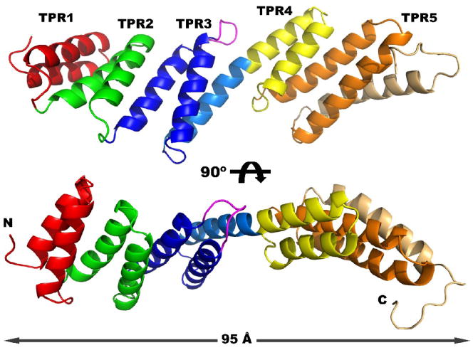
Figure 1. Crystal structure of rmBamD.
Two views of rmBamD related by a 90º rotation. The N-terminal domain contains TPRs 1–3 (red, green and blue respectively) and is capped by a helix (light blue). The C-terminal domain contains TPRs 4 and 5 (yellow and orange) and the final capping helix (light orange). An extended loop in TPR3 is labeled magenta.

