
Fig. 2.
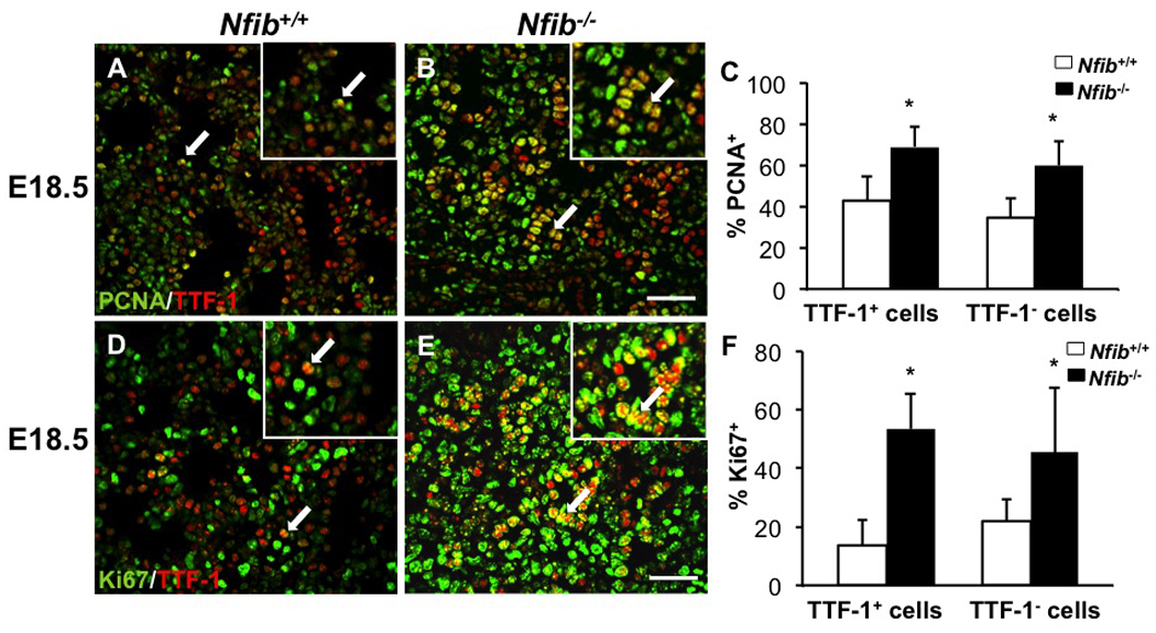
Loss of Nfib increases both epithelial (TTF-1+) and mesenchymal (TTF-1−) cell proliferation. Paraffin sections of E18.5 lungs from Nfib+/+ and Nfib−/− embryos were stained for both PCNA and TTF-1 (A, B) or for both Ki67 and TTF-1 (D, E). Immunostaining reveals there are more PCNA+TTF-1+ cells (arrows) and Ki67+TTF-1+ cells (arrows) in Nfib−/− lungs compared with Nfib+/+ lungs. Insets in panels A, B, D and E show a 5X magnification. Quantification (C, F) indicates there are more PCNA+TTF-1− cells and Ki67+TTF-1− cells in Nfib−/− lungs compared with Nfib+/+ lungs. Arrows denote the colocalization of PCNA and TTF-1 or Ki67 and TTF-1 and the region of magnification; *P<0.05; Scale bars, 50 µm.