
Fig. 6.
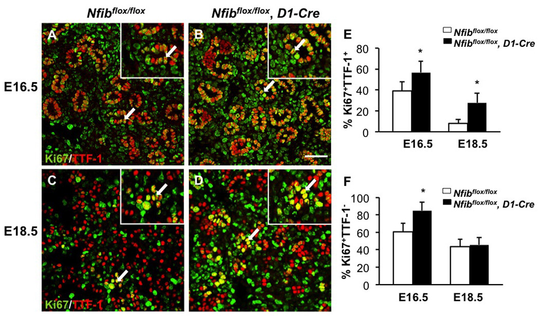
Loss of Nfib in mesenchyme increases TTF-1 positive and TTF-1 negative cell proliferation. Paraffin sections of E18.5 and E16.5 lungs from Nfibflox/flox and Nfibflox/flox, D1-Cre embryos were stained for both Ki67 and TTF-1 (A, B, C, D). Insets show a 5X magnification. Immunofluorescence staining reveals there were more Ki67+TTF-1+ cells (arrows) in Nfibflox/flox, D1-Cre lungs compared with Nfibflox/flox lungs at E16.5 and E18.5 (E). The percentage of Ki67+TTF-1− cells was also higher in Nfibflox/flox, D1-Cre lungs at E16.5 (F). In contrast, the percentage of Ki67+TTF-1− cells was similar between Nfibflox/flox and Nfibflox/flox, D1-Cre lungs at E18.5 (F). Arrows denote the colocalization of Ki67 and TTF-1 and the region of magnification; *P<0.05; Scale bar, 50 µm.