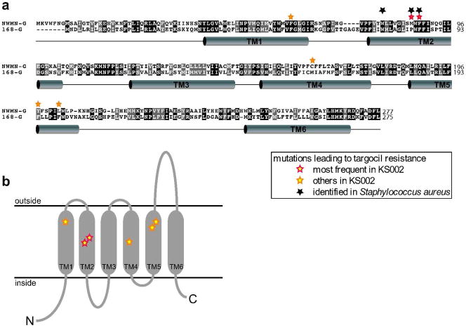
Figure 2. Alignment of B. subtilis and S. aureus TagG/TarG.
(a) Alignment of S. aureus TarG (NWMN-G) and B. subtilis TagG (168-G). Conserved amino acids are shown with a black background, similar amino acids with a grey background. Barrels indicate predicted transmembrane helices. b) Predicted topology of TarGSa with stars marking the sites of point mutations leading to targocil resistance in B. subtilis KS002.

