
Fig. 1.
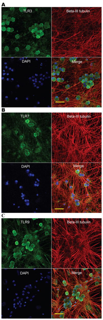
DRGNs express TLR3, TLR7 and TLR9. Confocal images of primary cultured mouse DRGNs isolated from E13 embryos are shown. DRGNs were stained with TLR3 (A), TLR7 (B) and TLR9 (C) (green), neuron-specific beta-III tubulin (red) and DAPI (blue). Shown are separate monochrome images of the green, red and blue fluorescence channel, and merged color images from all channels. Scale bars: 50 μm.