
Fig. 2.
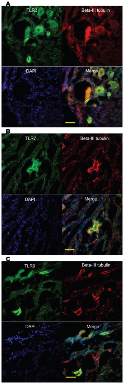
Human DRG sections express TLR3, TLR7 and TLR9. Confocal images of DRG cryostat sections. Samples were stained with TLR3 (A), TLR7 (B) and TLR9 (C) (green), neuron-specific beta-III tubulin (red) and DAPI (blue). Shown are separate monochrome images of the green, red and blue fluorescence channel, and merged color images from all channels. Scale bars: 50 μm.