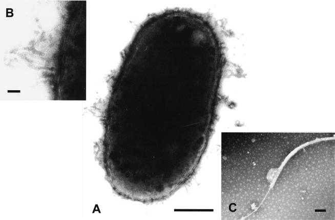
FIG. 1.
(A) Transmission electron photomicrograph of a 1% phosphotungstic acid negatively stained preparation of Vibrio fluvialis-like strain 1AMA showing typical Vibrio-like ultrastructure. Bar, 0.5 μm. (B) A higher magnification of a tubular structure typically seen in these organisms. Bar, 0.5 μm. (C) The single polar ensheathed flagellum. Bar, 0.1 μm.

