
Fig. 3.
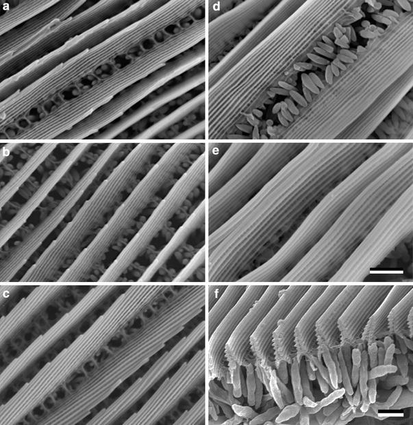
Scanning electron micrographs of single scales with structural colouration. a G. cleopatra forewing, b G. cleopatra hindwing, c G. rhamni, d H. glaucippe, e C. regina, f Sectioned scales of C. regina showing that the ridges are folded into a multilayer. A layer with large pigment granules exists beneath the ridges. In a–c the ridges were flexed to show the side-view of the lamellar stack (bars a–e, f 1 μm)