Fig. 3.
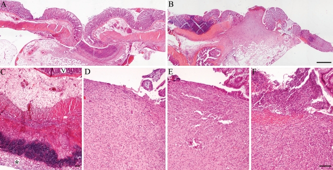
Light microscopy of anastomoses from different groups. A control anastomosis at 3 and 7 days follow-up (a and b, respectively). At 3 days follow-up, a collagen fleece (asterisk) induced an intense inflammatory reaction (c, v = villi). At 7 days follow-up, the control (d) and the fibrin glue (e) groups showed comparable wound healing at the luminal interface. In the collagen group (f), a more severe inflammation was still present (H&E staining, bars represent 500 μm (a and b, original magnification ×40) and 100 μm (c–f, original magnification ×100))

