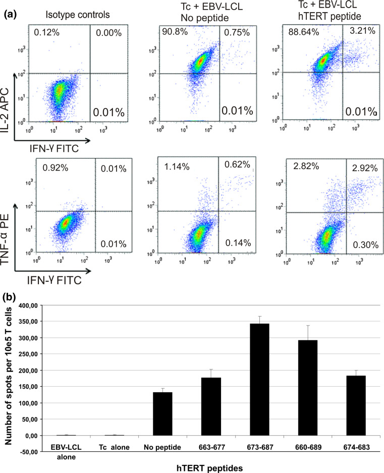
Fig. 6.
CD4+ T-cell cytokine secretion upon stimulation with a hTERT 30-mer peptide 660–689. PBMCs were cultured for 12 days in the presence of a hTERT peptide 660–689 containing several hTERT epitopes inducing T-cell proliferative responses. a The T-cell lines obtained from blood samples harvested 26 months post-vaccination start were restimulated overnight with autologous EBV-LCL loaded or not with peptide prior to intracellular cytokine staining. T cells were stained with CD4-PE-Cy7 or CD8-PB and IFN-γ-FITC, IL-2-APC and TNF-α-PE. Plots show cells gated on CD4+ T cells. b Frequency of peptide-specific IFN-γ secretion detected in ELISPOT assays from month 30 after start of vaccination. Autologous PBMCs were used as target cells. The E:T ratio was 2:1. Responses against peptides 663–667, 673–687, 660–689 and 674–683 were tested

