
Figure 1. Expression of Syb1 and Syb2 in WT and Syb1lew/lew mice.
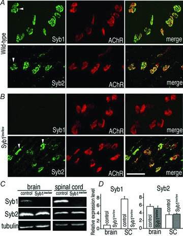
A and B, whole mounts of triangularis sterni (P14) muscles from WT mice (A) or Syb1lew/lew mutant mice (B) were immunostained with antibodies against Syb1 or Syb2, as well as Texas Red-conjugated α-bgt, which labels postsynaptic AChRs. Both Syb1 and Syb2 were detected at the WT nerve terminals (arrowheads in A). Syb1 was absent from Syb1lew/lew NMJs: only α-bgt staining was detected in the synaptic regions of the muscle (upper row in B). In contrast, Syb2 was detected in the nerve terminals (arrowheads in B) of Syb1lew/lew mutant mice. C, images of quantitative Western blot analysis of the brain and spinal cord (SC) from Syb1lew/lew and their littermate control mice (P14). D, comparison of relative levels of Syb1 or Syb2 between Syb1lew/lew mice (n = 3) and their littermates (control, n = 3). Relative expression levels of Syb1 and Syb2 were calculated by normalizing to that of α-tubulin (loading control). Syb1 was absent from Syb1lew/lew mice, but present in control mice, with significantly higher (P < 0.001) expression in the SC (7.73 ± 0.66, n = 3) compared to the brain (0.96 ± 0.21, n = 3) (normalized to α-tubulin level). In both Syb1lew/lew and control mice, Syb2 was more abundantly expressed in the brain compared to the SC (control: brain, 5.69 ± 0.57; SC, 3.54 ± 0.5, P < 0.05; Syb1lew/lew mutant: brain, 5.22 ± 0.57, SC, 3.69 ± 0.27, P < 0.05). In both the brain and SC, the relative levels of Syb2 remained similar between the Syb1lew/lew and control mice, P > 0.05. Scale bar in A and B, 50 μm.