
Figure 2. Formation of neuromuscular synapses in Syb1lew/lew mice.
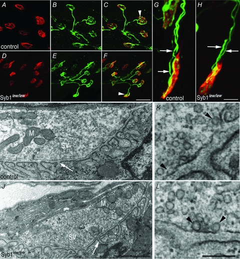
A–H, diaphragm muscles (P14) of control (A–C and G) and Syb1lew/lew mice (D–F and H) were doubly labelled with presynaptic markers (anti-synaptotagmin 2 and anti-neurofilament) (green) and postsynaptic markers (Texas Red-conjugated α-bgt) (red). In both control and Syb1lew/lew mice, nerve terminals (B and E) formed close apposition with postsynaptic AChRs (A and D), as shown in the merged images (white arrowheads in C and F). The majority (99%) of the end-plates were innervated by a single axon, except a small number (1%) of end-plates which were occupied by two axons in both control (G, white arrows point to two individual axons) and Syb1lew/lew mice (H, white arrows). I–L, electron micrographs of longitudinal sections of P14 diaphragm muscles. A typical nerve terminal from control (I) and Syb1lew/lew mice (J): both nerve terminals were packed with abundant synaptic vesicles (SV) and mitochondria (M). Postsynaptic junctional folds (white arrow in I and J) were morphologically normal in Syb1lew/lew mice compared with the control. K and L, examples of docked vesicles (black arrowheads) viewed under high magnification. K: control; L: Syb1lew/lew. Scale bars: A–F, 20 μm; G and H, 5 μm; I and J, 1 μm; K and L, 0.2 μm.