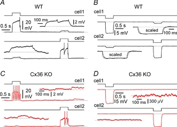
Figure 1. Electrical coupling between CA1 stratum lacunosum-moleculare interneurons in wild-type (WT, black traces) and connexin36 knockout (Cx36 KO, red traces) mice.

A, sequential injection of a depolarizing current step (90 pA, black steps) to interneurons of WT mice generates an attenuated response in the non-injected cell. Insets show the attenuated responses at enhanced magnification: notice the presence of spikelets. B, similar protocol as in A, but hyperpolarizing current steps of –50 pA (black steps) are injected. Notice the propagation of the voltage response to the non-injected neuron. Also notice, in the insets, after scaling, the slower kinetics of the propagated response (dotted line) vs. the original one (continuous trace). C and D illustrate the same type of experiments as A and B, respectively, performed on interneurons in a slice obtained from a Cx36 KO mouse. Notice the lack of a propagated response in the non-injected neuron. Current pulses were 50 pA in C (black steps) and –50 pA in D (black steps). Insets in C and D show the lack of a voltage response in the non-injected cell at a magnified scale. Gabazine (12.5 μm), NBQX (20 μm), d-AP5 (50 μm) and CGP55845 (1–5 μm) present throughout in A–D.
