
Figure 6. M2R structure and topology.
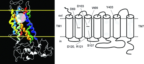
Left, homology model of M2R based on the rhodopsin crystal structure (Pedretti et al. 2006) showing the helical bundle formed by the 7 transmembrane helices. Border of cell membrane is delineated by the yellow lines. Orthosteric binding site is depicted by transparent circle. Right, topology of M2R showing positions of key amino acid residues that were mutated in this study.