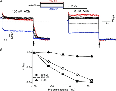
Figure 2. IKACh relaxation elicited by ACh in feline atrial myocytes.

A, macroscopic currents were induced by 100 nm (left) and 3 μm ACh (right) obtained with a double-pulse protocol (inset above). Currents at −120 mV were recorded after 3.5 s pre-pulses between −100 and +60 mV in steps of 40 mV. Current magnitude at the arrow is a measure of the number of KACh channels activated during the pre-pulse. Zero current level is indicated by the horizontal dashed line. B, relationship between the pre-pulse voltage and I/Imax ratio for currents induced by three ACh concentrations (n = 4–5). Current magnitude measured at arrow was normalized to the maximum current elicited by pre-pulse to −100 mV. For sub-saturating ACh concentrations, the fraction of KACh channels activated during the pre-pulse decreased with stronger depolarizations.
