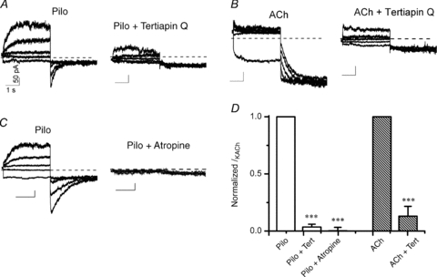
Figure 6. Atropine and tertiapin Q inhibit Pilo-activated currents in atrial myocytes.

Macroscopic current recordings from an isolated atrial myocyte activated by 3 μm Pilo (A) and 30 nm ACh (B) before (left) and during perfusion with 300 nm tertiapin Q (right). Currents were elicited by 3.5 s pre-pulses between −100 and +60 mV in steps of 40 mV, followed by step hyperpolarization to −120 mV. Vh was −40 mV. C, 3 μm Pilo-activated current before (left) and after perfusion with 100 nm atropine (right). The x–y scale bars indicate 1 s and 50 pA, respectively, for all current traces in A–C. D, fraction of IKACh inhibited by tertiapin Q or atropine. Current at the end of the 3.5 s voltage to +60 mV (Pilo) and −100 mV (ACh) after tertiapin Q or atropine treatment was normalized to pre-treatment value to obtain the relative amount of IKACh inhibited by tertiapin Q or atropine. n = 4 cells each group.
