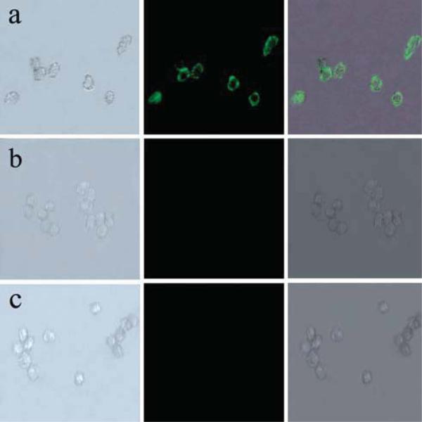
Fig. 8.
Images of live HeLa cells after being incubated with Fe3O4/NaYF4 nanocomposites biolabeled with transferrin (a), after being incubated with Fe3O4/NaYF4 nanocomposites without transferrin conjugated (b), and without incubating with any nanoparticles (c). In the three panels, the left rows are images in bright field, the central rows represent fluorescent images in dark field and the right rows are the overlays of the left and central rows.

