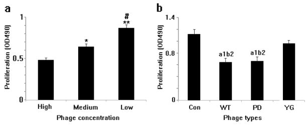
Figure 5. Proliferation of MSCs on the phage films derived from phages with different concentration (a) and surface chemistry (b).
MSCs cultured at the low concentration (1012 pfu/ml) of phage film grew faster than those at the higher concentration (1014pfu/ml) of film (a). MSCs seeded on the phage film grew slower than those on the glass slides (b). The growth rate of cells on the YG-phage film is faster than those on the WT- or PD-phage film. CON: glass slide coated with polylysine as control; YG: phage with osteogenic peptide displayed on the side wall; WT: wild type phage; PD: phage with osteocalcine-derived peptide (PDPLEPRREVCE) displayed on the side wall. (when compared with High group, *P<0.05, **P<0.01; compared with Medium group, #P<0.01; compared with control group, a1P<0.01; compared with YG group, b2P<0.05).

