Figure 3.
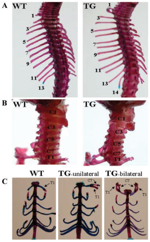
Abnormalities in the anterior/posterior axial skeleton of transgenic mice. Skeletons from 10-day-old mice were stained with alizarin red and alcian blue. A: Dorsal view of the anterior/posterior axial skeleton in wild-type (WT) and transgenic (TG) mice, showing TG mice have 14 pairs of ribs while WT mice have 13 pairs of ribs. B: Lateral view of cervical vertebrae in WT and TG mice. A supernumerary rib in TG mice is formed on the seventh cervical vertebra (C7). This cervical rib is fused to the middle of the rib that is articulated with the first thoracic vertebra (T1) and is attached to the sternum together at the T1 position in TG mice. C: Ventral view of vertebrosternal ribs in WT and TG mice. The cervical rib (C7 rib), which is fused to the T1 rib. In some cases it is unilateral (TG-unilateral) while it is bilateral (TG-bilateral) in other cases.

