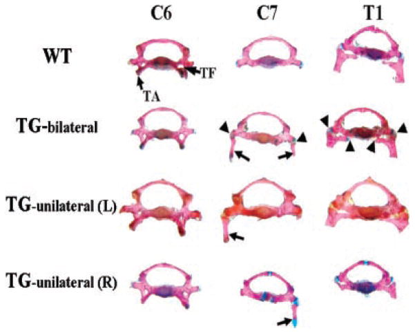
Figure 4.
Morphological appearances of the seventh cervical vertebra in transgenic mice. Vertebrae were dissected from alizarin red and alcian blue-stained skeleton. In wild-type (WT) mice, the C7 vertebral body resembles the C6 vertebral body instead of the T1 vertebral body without the tuberculi anterior (TA) and the transverse foramen (TF) that mark the C6. In transgenic mice there are ribs on C7, and the C7 vertebral body resembles the T1 vertebral body instead of the C6 vertebral body. This is more obvious in TG-bilateral mice. In TG-bilateral, TG-unilateral (L) and TG-unilateral (R) mice, there is only one joining point between the cervical ribs (indicated by arrows) and the C7 vertebral body while there are two joining points between the T1 ribs and the T1 vertebral body. The joining points between ribs and vertebral bodies are indicated with arrowheads only in TG bilateral but not in TG-unilateral (L) and TG-unilateral (R) mice. TG-bilateral =-transgenic mice showing C7 ribs on both sides; TG-unilateral (L) = transgenic mice showing C7 rib on the left side; TG-unilateral (R) = transgenic mice showing C7 rib on the right side.

