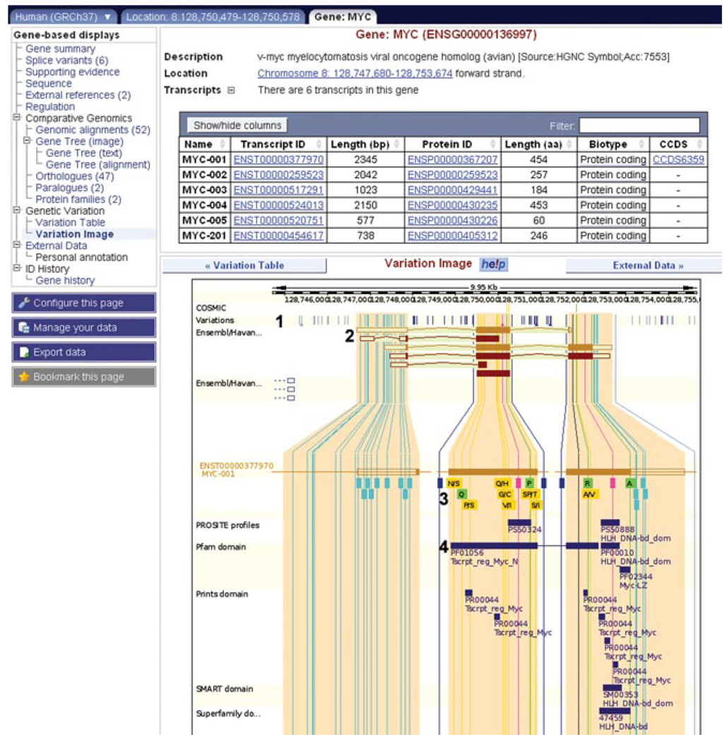
Figure 6.11.14.
The “variation image” in the gene tab. (1) All variations in MYC transcripts are displayed as vertical lines, color-coded according to the legend at the bottom of the view (not shown in the figure). (2) The six MYC transcripts are drawn. (3) The MYC-001 transcript is drawn, along with variations. Synonymous and nonsynonymous SNPs show encoded amino acid(s) in single letter code. For example, the yellow box showing “N/S” reveals that asparagine or serine can be coded for at that position. Clicking on a variation will open an information box, and a link to the variation tab. (4) Protein domains from various sources are drawn along the transcripts. For example, a transcript regulation domain in Pfam maps to the second and third exons of MYC-001. For color version of this figure go to http://www.currentprotocols.com/protocol/hg0611.

