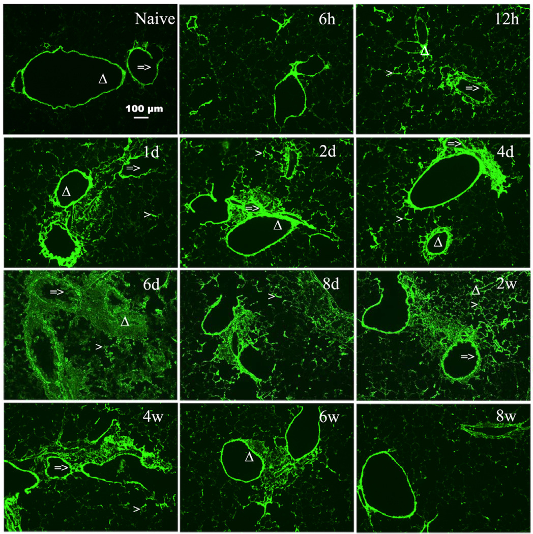
Figure 1. Increased HA accumulation in OCT sections from lungs isolated during acute and chronic stages of antigen exposure.
Sections were stained with the HABP as described in Methods (magnification bar 100 µm). HA deposition is shown for lungs from naïve (unchallenged) mouse and from OVA-sensitized mice during the course of allergen challenge. Mice were sacrificed at the following time points during the course of antigen exposure at 6 hours (6h), 12 hours (12h), 1 day (1d), 2 days (2d), 4 days (4d), 6 days (6d), 8 days (8d), 2 weeks (2w), 4 weeks (4w), 6 weeks (6w), and 8 weeks (8w).
=> indicates the airway of lung section.
Δ indicates the blood vessel of lung section.
> indicates the alveolar interstitium of lung section

