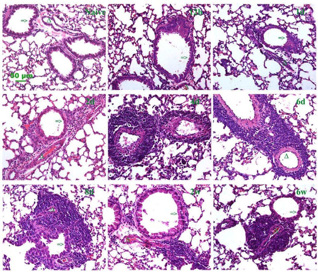
Figure 5. Histopathologic changes in lung sections during acute and chronic stages.
Hematoxylin and eosin (H&E) stained paraffin sections represent the inflammation infiltrate trend for the acute stage (12 h, 1 and 2 days) and chronic stage (4 days through 6 weeks) of antigen exposure. Clumping cells demonstrate the progression of perivascular and peribronchial pulmonary inflammation (magnification bar 50 µm). A section from a control lung (naïve) is also shown. Additional low magnification fields are shown in supplement figure 1. Mice were sacrificed at the time points during the course of antigen exposure as in figure 1.
=> indicates the airway of lung section.
Δ indicates the blood vessel of lung section.

