
Figure 1.
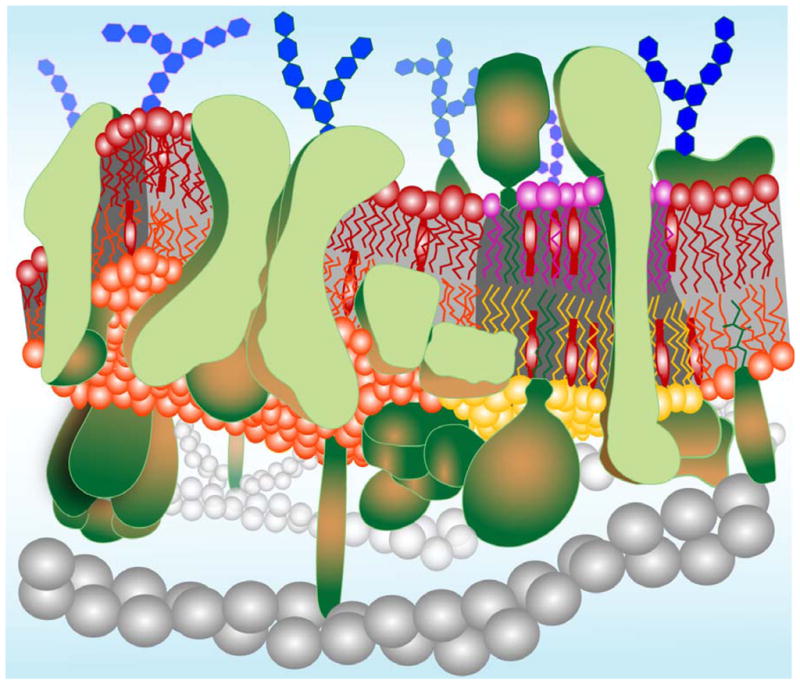
A contemporary model of a biological membrane. Lipids are organized in a dynamic bimolecular film with asymmetry across the membrane and a lateral organization in cholesterol-rich and cholesterol-poor domains. The outer leaflet (purple and red) is enriched in sphingomyelin and the inner leaflet (orange and yellow) is enriched in phosphatidylethanolamine, phosphatidylserine, and phosphatidylinositol. Both leaflets contain similar amounts of phosphatidylcholine and cholesterol (lollipop-shaped red structures). Cholesterol-rich lipid domains (“rafts”) are shown in register in both leaflets as determined experimentally (see text). Transmembrane, monotopic (partially inserted), and lipid-anchored proteins (green) occupy much of the total available membrane area and are also clustered into functional complexes in many cases. Glycosylations on proteins and lipids of the outer leaflet are shown as blue hexagon-shaped structures. Gray beaded helical structures symbolize elements of the cytoskeleton, which interact with proteins on the cytoplasmic leaflet of the membrane.