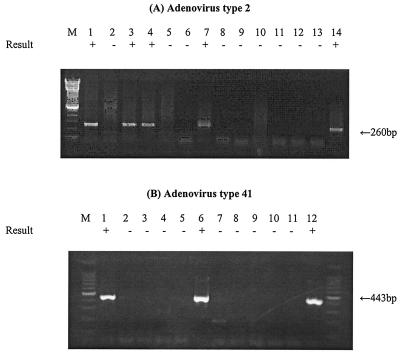
FIG. 5.
RT-PCR assay detection of Ad mRNA from cells inoculated with environmental water concentrates seeded with Ads. mRNA was recovered from infected cells after 5 to 7 days of incubation. (A) Lanes: 1 and 2, RT-PCR assay of Ad2 mRNA from A549 cells infected with 20 and 2 IU in North Carolina water concentrate; 3 and 4, RT-PCR assay of Ad2 mRNA from cells infected with 20 and 2 IU in Georgia water concentrate; 5, RT-PCR assay of mRNA in noninfected A549 cells; 6, negative control; 7, positive control; 8 and 9, PCR assay of Ad2 mRNA from A549 cells infected with 20 and 2 IU in North Carolina water concentrate; 10 to 11, PCR assay of Ad2 mRNA from cells infected with 20 and 2 IU in Georgia water concentrate; 12, PCR assay of mRNA in noninfected A549 cells; 13, negative control; 14, positive control. (B) Lanes: 1 to 3, RT-PCR assay of Ad41 mRNA from G293 cells infected with 10, 1, and 0.1 IU in Georgia water concentrate; 4, RT-PCR assay of mRNA in noninfected G293 cells; 5, negative control; 6, positive control; 7 to 9, PCR assay of Ad41 mRNA from G293 cells infected with 10, 1, and 0.1 IU in Georgia water concentrate; 10, PCR assay of mRNA in noninfected G293 cells; 11, negative control; 12, positive control. The detection sign (+ or −) was based on visual examination of the agarose gel for an amplicon of the correct size.

