Fig. 1.
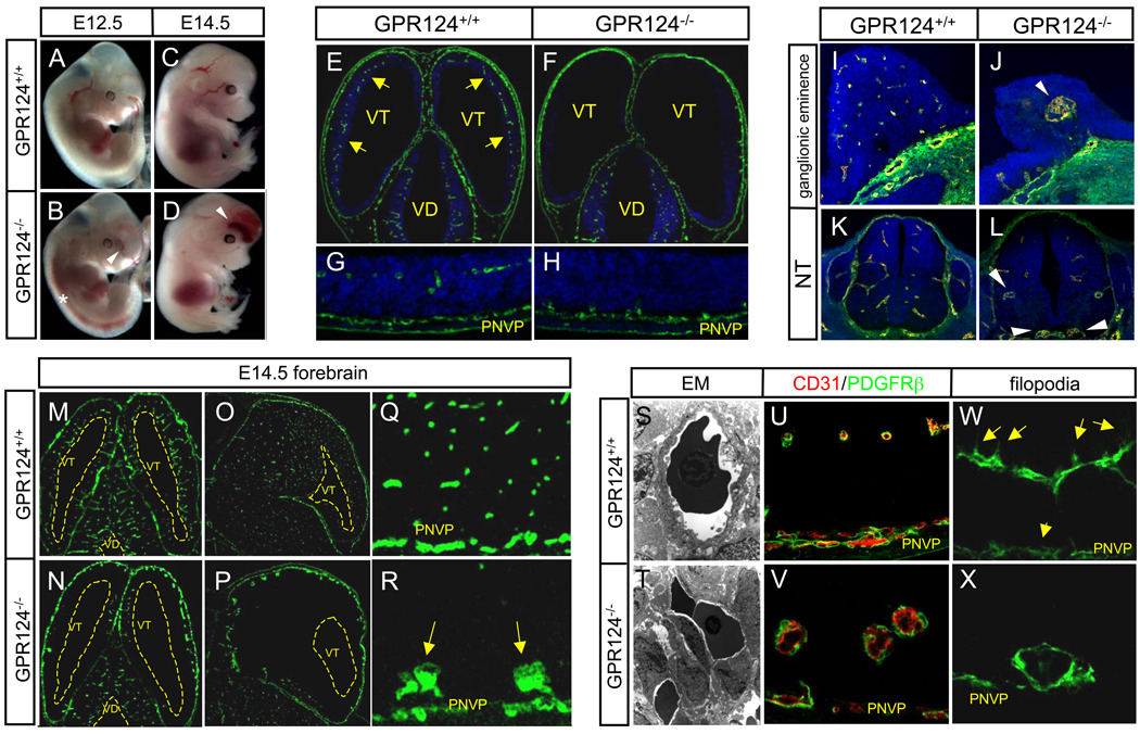
(A to X) CNS vascular patterning defects in GPR124−/− embryos. GPR124−/− embryos manifested progressive hemorrhage confined to forebrain telencephalon (arrowheads) and neural tube (*) [(A) to (D)]. Wild type E11.5 embryos exhibited efficient sprouting angiogenesis (arrows) from the perineural vascular plexus (PNVP) into the telencephalon [(E) and (G)], which was completely ablated in GPR124−/− embryos that exhibited PNVP thickening only [(F) to (H)], Green- CD31 IF. Large, glomeruloid vascular malformations (arrowheads) and absence of a vascular network were observed in E11.5 GPR124−/− forebrain ganglionic eminences and ventral neural tube [(I) to (L), CD31 IF]. By E14.5, GPR124−/− telencephalon was essentially devoid of a mature capillary network with glomeruloid malformations abutting the pia/PNVP, while the diencephalon was unaffected [(M) to (N), transverse; (O) to (P), sagittal; vascular malformations (arrows), (Q) and (R), CD31 IF]. Glomeruloid malformations were irregular, multi-layer endothelial aggregates versus single layer capillaries in controls [(S) and (T), EM 4300x], with normal peripheral PDGFRβ+ pericyte investment [(U) and (V)] but lacked ventricularly-directed endothelial filopodia (arrows) [(W) and (X), CD31 IF]; VD, ventricle diencephalons; VT, ventricle telencephalon; blue, DAPI.

