
Fig. 7.
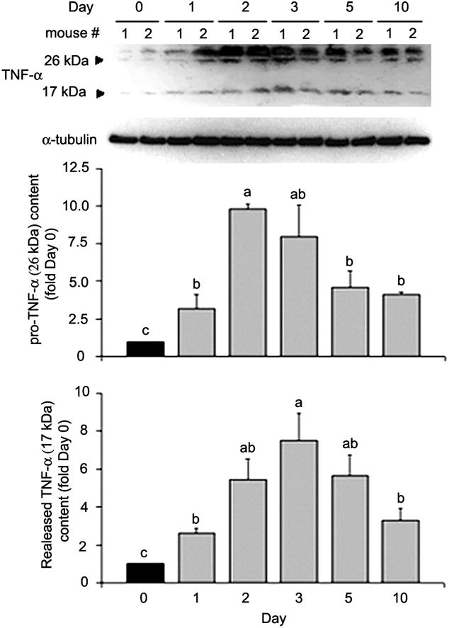
TNF-α release is increased in cardiotoxin-injured muscle. Cardiotoxin was injected into the soleus muscle of male adult mice (8 wk of age). Solei were collected before (day 0) or after injection at indicated times. Muscle lysates were analyzed by Western blot analysis with antibody against TNF-α, with antibody against α-tubulin as loading control. Optical density of TNF-α detected was normalized to α-tubulin and analyzed by ANOVA (P < 0.05 for both forms of TNF-α). Data were analyzed and expressed as described in Fig. 4; “ab” denotes that the value is not different from either a or b.