Figure 1.
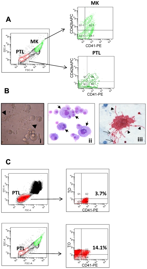
Phenotypical characterization of human primary MK cultures. (A) Flow cytometric analysis of MK cultures labeled with PE-conjugated anti-CD41 and APC-conjugated anti-CD42 antibodies. The density plot on the left demonstrates the forward scatter and side scatter properties of the cells that were acquired on a logarithmic scale to identify both MKs and platelet-sized cells (PTL). The density plots on the right show CD41+/CD42b− and CD41+/CD42b+ MKs and culture-derived PTL present in the bottom right and top right quadrants, respectively. (B) MKs derived from CD34+ hematopoietic progenitor cells were visualized by phase-contrast (i) and light microscopy after Wright-Giemsa (ii) and GPIIb/IIIa staining (iii). Mature MKs with polylobulated nuclei and large proplatelet-bearing MKs are present in liquid culture (i-ii, arrows); MKs grown in semisolid culture display cytoplasmic extensions or proplatelets (iii, arrowheads) with nascent platelets at their ends (iii, arrows). (C) Flow cytometric analysis of culture-derived platelets labeled with anti-CD41 antibodies and TO. The dot plots in the upper panels represent human PB PTL labeled with CD41 antibodies (x-axis) and TO (y-axis), which served as a positive control. The plots in the bottom panels represent culture-derived PTL labeled in the same manner.

