Figure 3.
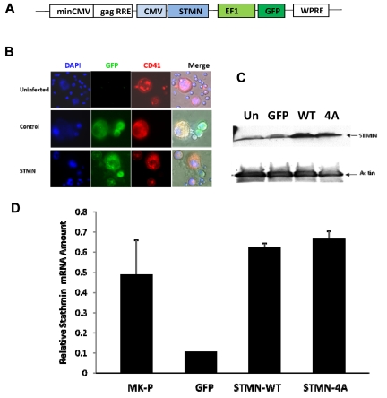
Lentivirus-mediated delivery of stathmin in mature MKs. (A) Schematic representation of the transfer vector backbone of the FIV-based lentiviral system in which stathmin (STMN) cDNA is driven by cytomegalovirus (CMV) promoter and GFP gene is driven by elongation factor 1 (EF1) promoter. (B) Immunofluorescence microscopy analysis of MKs infected with stathmin-expressing lentiviruses (green fluorescence) that were labeled with PE-conjugated anti-CD41 antibodies (red fluorescence) and with Hoechst 33342 to visualize DNA and nuclear morphology (blue fluorescence). Image acquisition was performed using a Zeiss Axioplan 2 microscope equipped with a 40×/0.75 dry objective and an AxiCam MRm camera (Carl Zeiss). Image analysis was performed using Zeiss AxioVision LE software. (C) Western blot analysis of protein lysates extracted from 293HEK cells uninfected (Un), infected with control (GFP), and WT or mutant (4A) stathmin-expressing lentiviruses. After blotting, the membranes were incubated with anti–human stathmin polyclonal antibody (top panel) and with antiactin monoclonal antibodies, the latter used as control for protein loading. (D) Quantitative real-time PCR analysis of mRNA extracted from MKs infected with control (MK + GFP), wild-type (MK + STMN-WT), or mutant (MK + STMN-4A) stathmin lentiviruses. mRNA extracted from day 3 MK progenitors (MK-P) was also included in the analysis as reference for basal levels of stathmin expression. Each column represents the mean ± SD of 3 independent experiments performed in duplicates and normalized to GAPDH.

