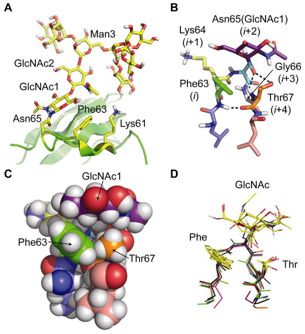
Figure 1.
(A) Ribbon diagram of the published NMR structure of HsCD2ad (PDB: 1GYA) (16). The N-glycan, Asn65, Phe63, and Lys61 are highlighted in yellow. (B) Stick and (C) space-filling representations of the type I β-bulge turn in HsCD2ad; the (i+2)-position Asn65(GlcNAc1) packs against the i-position Phe63, and the i+4 Thr67. Dashed lines indicate hydrogen bonds. (D) Glycosylated type I β-bulge turns with an i-position Phe from known proteins in the PDB (green: HsCD2ad; blue: 1G82; orange: 1FJR; red: 1ICF; and black: 3DMK). All structures rendered in Pymol (www.pymol.org).

