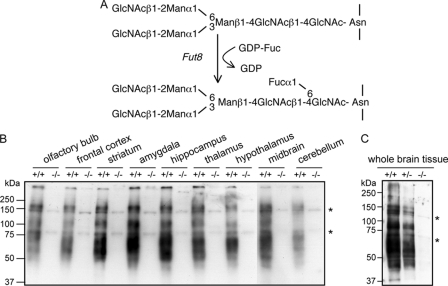
FIGURE 1.
Expression of α1,6-fucosylation in brain tissues. A, reaction pathway for the synthesis of α1,6-fucose. GlcNAc, N-acetylglucosamaine; Man, mannose; Fuc, fucose; GDP-Fuc, guanosinodiphospho-fucopyranoside. Homogenates of various brain regions from Fut8+/+ and Fut8−/− mice (B) and whole brain tissues from Fut8+/+, Fut8+/−, and Fut8−/− mice (3 months old) (C) were subjected to a lectin blot analysis using AAL as described under “Experimental Procedures.” Asterisk indicates nonspecific bands.

