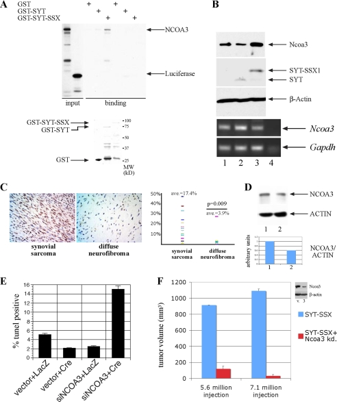
FIGURE 1.
SYT-SSX regulates NCOA3 protein. A, NCOA3 specifically interacts with SYT-SSX1. NCOA3 and luciferase as a control protein were radioactively labeled with [35S]methionine through in vitro translation, and were bound to GST, GST-SYT, and GST-SYT-SSX1. The bound proteins were washed and analyzed by SDS-PAGE. The input recombinant GST proteins used in these experiments were visualized through Coomassie staining shown in the lower panel. B, Ncoa3 protein and transcript level in rat1 3Y1 cells with stable expression of vector (1), SYT (2), and SYT-SSX1 (3) expression, were detected by immunoblot using antibody against NCOA3 and RT-PCR. SYT and SYT-SSX1 expression were detected with anti-Flag antibody (15). Lane 4 is a negative control without input. C, NCOA3 is significantly more expressed in synovial sarcomas compared with that in benign neurofibromas. Arbitrary staining intensity was measured by scoring methods outlined in methods section. The plot indicates the percentage of positive staining cells in the two tumor populations. Student's t test was applied with p value of 0.009. D, NCOA3 is decreased in SYO-1 cells treated with antisense oligonucleotide for SYT-SSX1/2. Lane 1: control oligonucleotide; lane 2: antisense oligonucleotide. The relative intensity of the NCOA3 and ACTIN level were quantitated by PhotoshopTM histogram analysis. E, rat1 3Y1cells with reduced Ncoa3 are more apoptotic in anchorage-independent growth. Cells infected with a conditional siRNA for Ncoa3 or a control construct was treated with Adeno-LacZ or Adeno-Cre to activate the siRNA expression for 2 days before cells were seeded into methylcellulose. After 48 h, cells were collected from methylcellulose, and the percentage of TUNEL-positive cells was quantitated through FACS analysis. F, decreased Ncoa3 expression results abrogation of tumor formation in vivo. SYT-SSX1-expressing rat fibroblast and those with reduced Ncoa3 expression (clone 3) were injected into both flanks of two nude mice, tumor formation was assessed 4 weeks later with measurement of tumor volume.

