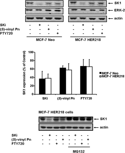
FIGURE 4.
Proteasomal degradation of SK1. MCF-7 Neo or MCF-7 HER2 cells were treated with SKi or FTY720, or (S)-FTY720 vinylphosphonate ((S)-vinyl Pn) (all at 10 μm final concentration) for 24 h. MCF-7 HER2 cells were also pretreated with MG132 (10 μm) for 30 min prior to addition of SK1 inhibitors. Western blots were probed with anti-SK1, anti-ERK-2, and anti-actin antibodies. Results are representative of three independent experiments. Bar graph shows quantification of the effect of the SK1 inhibitors on the proteasomal degradation of SK1 in MCF-7 Neo or MCF-7 HER2 cells by calculating the SK1:actin ratio for each treatment (p < 0.05 for control versus SK1 inhibitor-treated cells, n = 3 experiments).

