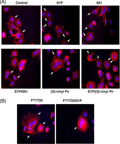
FIGURE 5.
Actin rearrangement in MCF-7 cells. MCF-7 Neo cells were treated with (A) SKi or (S)-FTY720 vinylphosphonate ((S)-vinyl Pn) or (B) FTY720 (both at 10 μm final concentration) for 15 min prior to stimulation with and without S1P (1 μm, 5 min). Actin was detected using phalloidin red staining. Arrows in the panel for S1P treatment identify actin localized to lamellipodia/membrane ruffles, whereas arrows in the other panels identify actin clustered into focal adhesions. Nuclei were stained with DAPI. Results are representative of three independent experiments.

