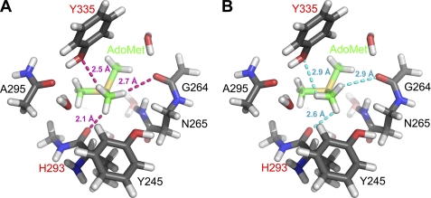
FIGURE 2.
Optimized active site with bound AdoMet (A) and manually rotated geometry (B) to eliminate CH···O hydrogen bonds. Truncated AdoMet and the protein are depicted with green and gray carbon atoms, respectively. Residues labeled in red designate CH···O acceptors. H···O distances from methyl protons to nearest oxygen atom for optimized and broken geometry are shown in magenta and cyan, respectively. The crystal structure of the SET7/9·AdoMet complex (Protein Data Bank code 1N6A) was used as the model for the calculations.

