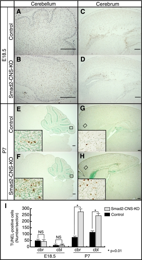
FIGURE 5.
Smad2-CNS-KO mice cerebella and cerebrum display more TUNEL-positive cells than those of control mice. A–H, sagittal brain sections of E18.5 and P7 mice were labeled for TUNEL. At E18.5, Smad2-CNS-KO mice cerebella (A and B) and cerebrum (C and D) showed a similar number of TUNEL-positive cells per section. At P7, compared with those of control mice, Smad2-CNS-KO mice cerebella (E and F) and cerebrum (G and H) showed a higher number of TUNEL-positive cells per section. Scale bar, 200 μm. Insets, high magnification images were taken from the areas indicated by rectangles. Scale bar, 50 μm. Arrowheads indicate TUNEL-positive cells. I, measurement of TUNEL-positive cells in E18.5 and P7 mice. Quantification revealed that at E18.5, no significant differences were found between Smad2-CNS-KO and control mice. However, the number of apoptotic cells is significantly higher in both Smad2-CNS-KO mice cerebellum and cerebrum at P7. Values are expressed as the mean ± S.D. per section of five sections per animal, three animals per group. NS, not significant. An asterisk represents p values < 0.01.

