FIGURE 4.
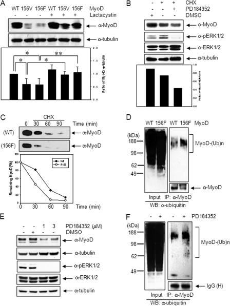
MyoD-Y156 phosphorylation is required for protein stabilization. A, 10T1/2 cells transfected with wild type MyoD or mutant MyoD (-Y156V (156V) or -Y156F (156F)) were kept in the absence or presence of lactacystin (10 μm), a proteasome inhibitor for 8 h, extracted, and analyzed by immunoblotting, using anti-MyoD (C-20) antibody. The ratio of MyoD protein to α-tubulin is shown in the lower panel. B, 10T1/2 cells were transiently transfected with MyoD expression plasmid. On the next day, the cells were pretreated with either PD184352 (1 μm), a specific MEK inhibitor for 30 min or not, and the protein synthesis was blocked by treatment of cycloheximide (CHX; 10 μg/ml), a protein synthesis inhibitor for an additional 30 min. The MyoD protein level was analyzed by immunoblotting using anti-MyoD (C-20) antibody. Reduction of pERK1/2 intensity showed that the MEK inhibitor were effective. C, 10T1/2 cells transfected with either wild type or mutant MyoD were treated with cycloheximide (10 μg/ml) for the indicated time. MyoD protein levels were analyzed by immunoblotting, using anti-MyoD (C-20) antibody. Quantitation of MyoD turnover following cycloheximide treatment is shown in the bottom panel. D, 10T1/2 cells transfected with either wild type or mutant MyoD-Y156F were treated with lactacystin (5 μm) for 12 h. Cell extracts were immunoprecipitated with anti-MyoD (C-20) antibody, and the polyubiquitination levels of endogenous MyoD were analyzed by immunoblotting, using anti-ubiquitin (Ub) antibody. E, C2C12 cells incubated in DM for 1 day were treated with PD184352 (1 or 3 μm) for 2 days, and the MyoD protein level was analyzed by immunoblotting using anti-MyoD (C-20) antibody. F, differentiating C2C12 myoblasts kept in DM for 1 day were treated with lactacystin (5 μm) for 12 h in the presence or absence of PD184352 (1 μm). Cell extracts were immunopreciptated with anti-MyoD (C-20) antibody, and the polyubiquitination levels of endogenous MyoD were analyzed by immunoblotting using anti-ubiquitin antibody. The results show the mean ± S.D. of triplicate experiments. *, p < 0.05; **, p < 0.01. WB, Western blot.

