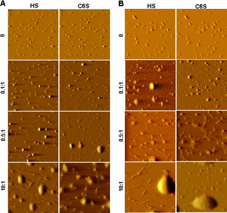
FIGURE 5.
Morphologies of R16 and K16 polyplexes treated with GAGs as seen by atomic force microscopy. Polyplexes of R16 (A) and K16 (B) were treated with increasing amounts of HS and C6S expressed as GAG:peptide w/w ratio. 2 μl of the resulting complex was deposited on mica, air-dried, and imaged in air with an atomic force microscope.

