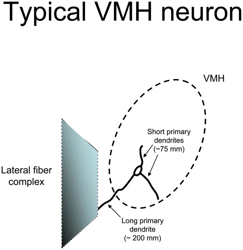
Figure 1.
Illustration of the dendritic arbour of a typical VMH neurone. The VMH is demarcated by dashed line forming an oval. The lateral fibre complex is portrayed as a shaded trapezoid. The typical VMH neurone is shown with three primary dendrites, with the longest primary dendrite extending towards the lateral fibre complex. The short primary dendrites remain within the borders of the VMH, presumably to be innervated by local interneurones.

