
Figure 1. Acute ischemic response has subsided 3-months post-injury.
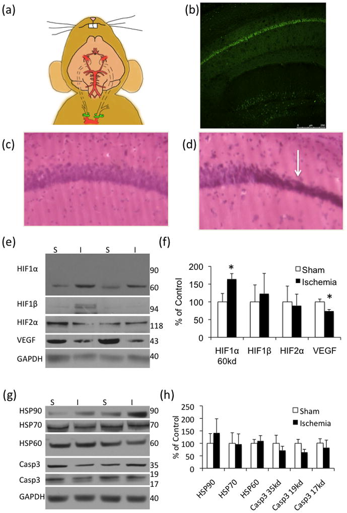
(a) Schematic of the bilateral occlusion of the common carotid artery utilized in the 3xTg-AD mice. Clamps are highlighted in green. (b) 24-hours post-ischemia there is abundant fluorojade staining in the hippocampus (n=4 sham, 4 ischemia). (c–d) H&E stain of sham and ischemic tissue, respectively. (e–f) Hypoxia inducible factors were mostly unchanged 3-months post-injury although vascular endothelial growth factor (VEGF) was significantly decreased in the ischemia-treated mice (n=12 sham, 14 ischemia). (g–h) Analysis by western blot of heat shock proteins (HSP) and caspase3 levels (casp) revealed no differences between ischemia-and sham-treated mice 3-months post injury (n=4 sham, 5 ischemia).