
Figure 4. Ischemia alters APP C-terminal fragments.
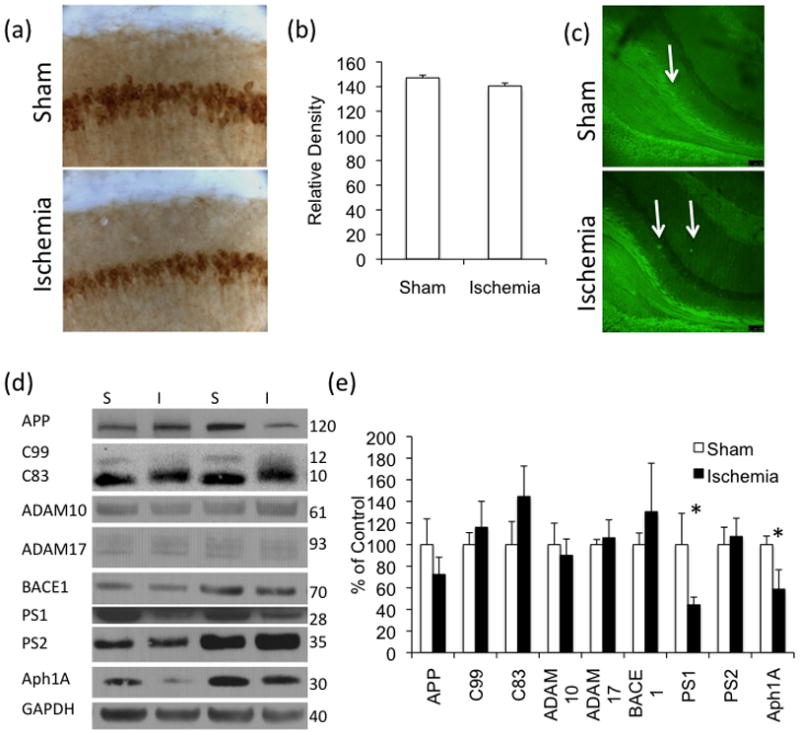
(a–b) Aβ-like staining, by 6E10 antibody, is not altered by immunohistochemistry 3-months post-ischemia. Quantification was performed using ImageJ and selecting an elliptical area of the CA1 (c) We did not find a difference in thioflavin staining. Arrows indicate areas of high thioflavin staining. (d–e) Western blot analysis of APP and APP-related proteins revealed a weight shift in the APP C-terminal fragment C83, but no changes in ADAM10, ADAM17 or BACE-1. We found a significant decrease in the γ-secretase components PS1 and Aph1 but not in PS2 (n=4 sham, 5 ischemia).