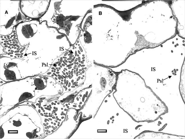
FIG. 3.
Transmission electron micrographs of ultrathin sections from cucumber leaves challenged with P. syringae pv. lachrymans at 96 h postchallenge. (A) Bacterial cell colonization of leaves sampled from nonelicited, challenged plants (T−Psl+). P. syringae pv. lachrymans progresses towards the inner leaf tissues mainly by intercellular (IS) growth. (B) Bacterial cell colonization of intercellular spaces in leaves of plants preelicited with T. asperellum 48 h prior to challenge (T+Psl+). Considerably fewer bacterial cells are observed. Bars, 2 μm.

