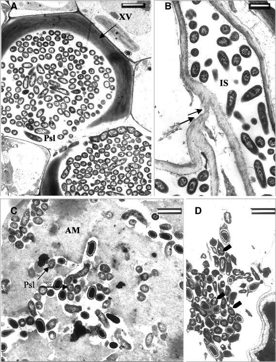
FIG. 4.
Transmission electron micrographs of ultrathin sections from cucumber leaf tissues. (A) Xylem vessels (XV) heavily colonized with P. syringae pv. lachrymans in leaves of nonelicited plants (T−Psl+) at 96 h postchallenge with P. syringae pv. lachrymans. (B) Digestion of host cell wall by P. syringae pv. lachrymans (arrows) in leaves of nonelicited (T−Psl+) plants. (C) Intercellular spaces (IS) of T. asperellum-preelicited plants (T+Psl+) in which an amorphous matrix (AM) has accumulated. P. syringae pv. lachrymans cells trapped in this AM appear embedded, and their development is halted. (D) P. syringae pv. lachrymans cells (arrows) in leaves of T. asperellum-preelicited plants (T+Psl+) appear to be severely damaged, and their content is aggregated. Some of these cells accumulate dark-staining osmiophilic material. Bars, 2 μm (A and D) and 1 μm (B and C).

