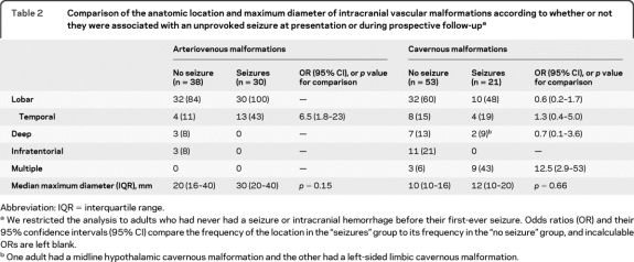
Table 2.
Comparison of the anatomic location and maximum diameter of intracranial vascular malformations according to whether or not they were associated with an unprovoked seizure at presentation or during prospective follow-upa
Abbreviation: IQR = interquartile range.
We restricted the analysis to adults who had never had a seizure or intracranial hemorrhage before their first-ever seizure. Odds ratios (OR) and their 95% confidence intervals (95% CI) compare the frequency of the location in the “seizures” group to its frequency in the “no seizure” group, and incalculable ORs are left blank.
One adult had a midline hypothalamic cavernous malformation and the other had a left-sided limbic cavernous malformation.

